


Saiful Islam: Laboratory Spot Check – Microcytic Anemias and The Thalassemia Spectrum
Saiful Islam, Lab TA 2 at Inova Fairfax Hospital, shared a post on LinkedIn:
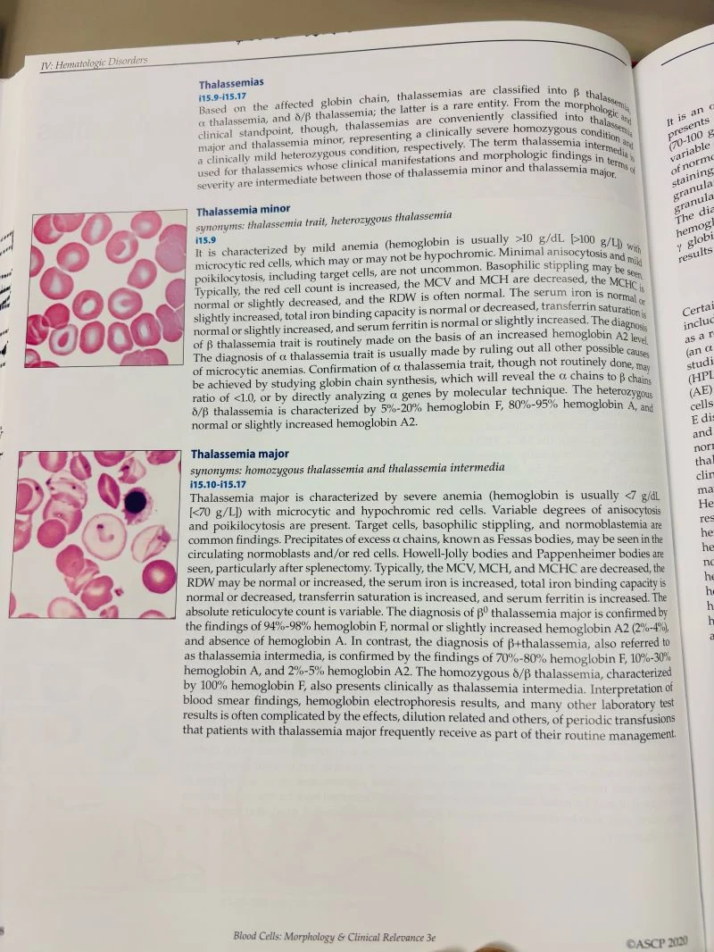
“Laboratory Spot Check: Microcytic Anemias and The Thalassemia Spectrum
In clinical hematology, distinguishing microcytic anemias requires looking beyond basic indices to assess peripheral blood morphology and hemoglobin electrophoresis profiles.
Here is a quick refresher on key differentiators when evaluating Thalassemia Minor vs. Thalassemia Major:
Thalassemia Minor (Trait)
- Presentation: Mild anemia (typically \text{Hb} > 10\text{ g/dL}), microcytosis, and mild hypochromia.
- Red Cell Count: Characteristically elevated relative to the degree of anemia, often alongside a normal RDW.
- Morphology: Target cells and basophilic stippling are commonly observed.
Key Diagnostic Findings:
- In \beta-thalassemia trait, elevated Hemoglobin A2 (\text{HbA}_2) levels are standard.
- In \alpha-thalassemia trait, confirmation relies on ruling out other causes of microcytic anemia, globin chain synthesis ratios (\alpha/\beta < 1.0), or molecular genetic testing.
Thalassemia Major
- Presentation: Severe microcytic, hypochromic anemia (frequently \text{Hb} < 7\text{ g/dL}).
- Morphology: Marked anisocytosis and poikilocytosis, target cells, basophilic stippling, and circulating normoblasts. In post-splenectomy patients, inclusions such as Howell-Jolly bodies, Pappenheimer bodies, and Fessas bodies (precipitated excess \alpha-chains) are frequently visualized.
Electrophoresis Profiles:
- /beta^0-thalassemia major: marked elevation of \text{HbF} (94\% \text{–} 98\%), absence of \text{HbA}.
- /beta^+-thalassemia: predominant \text{HbF} (70\% \text{–} 80\%) with residual \text{HbA} (10\% \text{–} 30\%).
A key diagnostic reminder for the lab: interpreting hemoglobin electrophoresis and smear morphology in severe thalassemias can be complicated by periodic transfusion history—always verify patient clinical history when reviewing unexpected profiles!
What red cell morphology clues do you find most distinct when differentiating microcytic anemias in your laboratory workflow?”
Stay updated with Hemostasis Today.
-
Oct 12, 2026, 07:39Emmanuel J Favaloro: 100 Years of von Willebrand Disease and the Importance of Local Diagnosis and Management
-
Oct 12, 2026, 07:39Post-Stroke Fatigue Trajectories – 38% Recovered at 12 Months, While 16% Developed New Fatigue – International Journal of Stroke
-
Oct 12, 2026, 07:28Beth Sara: IBMS Specialist Portfolio Achievement and a New Role in Transfusion Practice
-
Oct 12, 2026, 07:28Abdul Mannan: PCC vs. aPCC – The Small ‘a’ That Changes Emergency Management
-
Oct 12, 2026, 07:20Mokhtar Tazik: Improving Access to Plasma Derived Medicines Through Awareness, Interest and Accessibility
-
Oct 12, 2026, 07:08Larissa Chastain: Partnering to Transform Anticoagulation Management Through INR Self-Testing
-
Oct 12, 2026, 06:45Nick Lenamon: Stroke Awareness at Any Age Recognizing the Signs and Acting FAST
-
Oct 12, 2026, 04:45Aparna Korisetty: Upper GI Bleeding and Severe Anemia Secondary to Duodenal NET
-
Oct 12, 2026, 04:38Aya AlFakih: Why HbA1c Can Be High even When this Morning’s Glucose Looks Normal?