


Milad Abdalla: Decoding Hemoglobin Electrophoresis – A Practical Diagnostic Reference
Milad Abdalla, Medical Laboratory Technologist at Al Yalayis Government Transactions Center, shared a post on LinkedIn:
“Decoding Hemoglobin Electrophoresis: A Practical Diagnostic Reference
Accurate interpretation of hemoglobin fractions is essential for identifying inherited hemoglobin disorders and differentiating thalassemia syndromes.
Hemoglobin electrophoresis and complementary techniques, such as HPLC and capillary electrophoresis, help identify abnormal hemoglobin patterns and quantify major fractions.
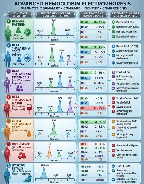
Here is a practical overview of the characteristic patterns:
1. Normal Adult Hemoglobin Pattern
- HbA (HbA₀): approximately 95–98%
- HbA₂: approximately 2.0–3.5%
- HbF: usually less than 1%
2. Beta-Thalassemia Trait (Minor)
- Increased HbA₂ (typically 3.5% or more).
- HbA is usually the predominant fraction.
- HbF may be mildly elevated.
- Commonly associated with microcytosis and hypochromia.
3. Beta-Thalassemia Intermedia and Major
- Reduced or absent HbA, depending on the genotype.
- Significantly increased HbF.
- HbA₂ may be elevated or variable.
- In transfusion-dependent beta-thalassemia, HbF can represent a substantial proportion of total hemoglobin.
4. Alpha-Thalassemia Spectrum
- Silent Carrier or Alpha-Thalassemia Trait: Often normal adult hemoglobin fractions; genetic testing may be required for confirmation.
- HbH Disease: Presence of HbH (β₄), often approximately 5–30%, depending on disease severity.
- Hb Bart’s Hydrops Fetalis: Predominance of Hb Bart’s (γ₄), frequently exceeding 80% in severe cases.
Important Laboratory Considerations
Hemoglobin fractionation should never be interpreted in isolation.
Always correlate the results with:
- CBC indices (MCV, MCH, RBC count).
- Iron studies, particularly ferritin.
- Patient age and clinical history.
- Previous blood transfusions.
- Family history and molecular testing when indicated.
A key diagnostic challenge: Iron deficiency may coexist with beta-thalassemia trait and potentially influence HbA₂ interpretation.
Therefore, borderline results require careful clinical and laboratory correlation.”
Stay updated with Hemostasis Today.
-
Nov 10, 2026, 16:28Can the VWF to ADAMTS-13 Ratio Predict Mortality in Sepsis-Induced Coagulopathy? – RPTH Journal
-
Nov 10, 2026, 16:28Ruth Willems: PhD Defence at Maastricht University on Thrombosis and Bleeding in HPB Disease
-
Nov 10, 2026, 16:09Edward Lee Carter: Factor XIa Inhibition Enters Clinical Practice
-
Nov 10, 2026, 16:08Edward C.C. Wong: Deriving Ferritin Cutoffs for Iron Deficiency Using RBC Parameters
-
Nov 10, 2026, 15:13Wolfgang Miesbach: Nonviral Gene Therapy for Haemophilia A Using Lipid Nanoparticles
-
Nov 10, 2026, 15:06Dahra Teles: First Speaking Experience at HEMO 2026 on First-Line Multiple Myeloma Treatment
-
Nov 10, 2026, 14:55Yazan Abou-Ismail: First FDA-Approved Factor XIa Inhibitor for Stroke Prevention
-
Nov 10, 2026, 14:49Ifeanyichukwu Ifechidere: The Role of Coagulation in Anaesthesia and Surgery
-
Nov 10, 2026, 14:37Salih Ehsan: Common Causes of Microcytic Anemia