


Hassan Abdelhak: Understanding How LVADs Support the Failing Heart
Hassan Abdelhak, L-CRN CCL at King Salman Specialist Hospital, Taif, shared a post on LinkedIn:
“Understanding Left Ventricular Assist Devices (LVAD)
When advanced heart failure reaches stage D, traditional medical therapy often hits a wall. For patients awaiting a transplant – or those who aren’t candidates for one – a Left Ventricular Assist Device (LVAD) serves as a mechanical lifeline.
Understanding how an LVAD works!?
How an LVAD works: step-by-step
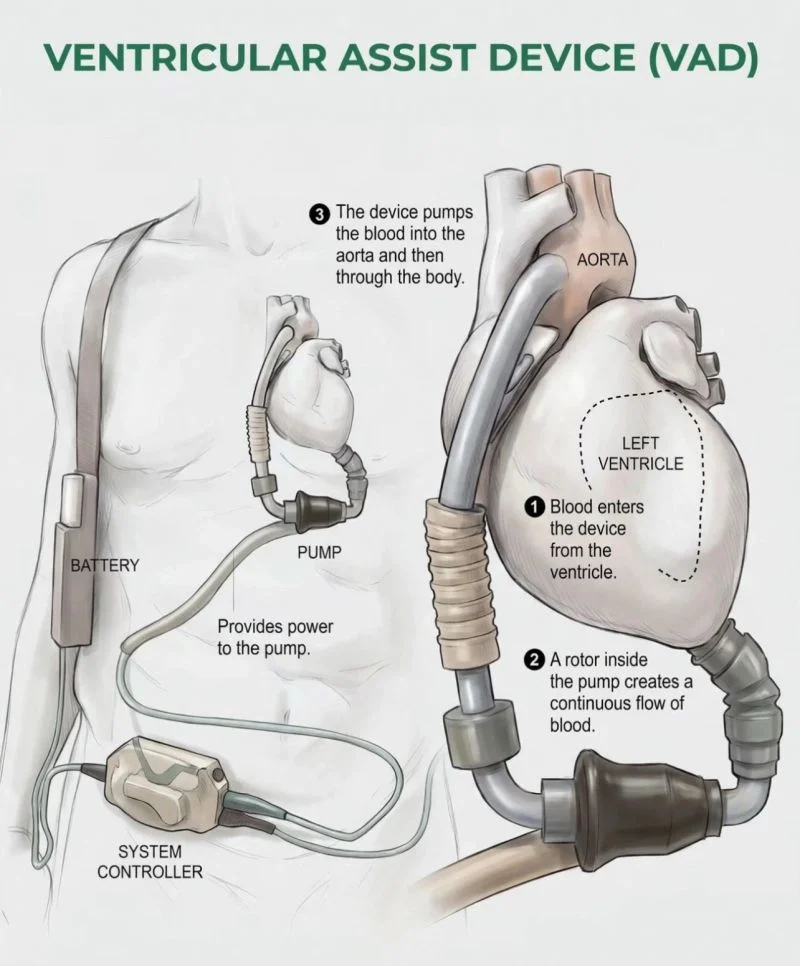
As shown in the diagram:
- Inflow (blood draw): Blood enters the mechanical pump directly from the left ventricle apex, bypassing the weakened native myocardium.
- Continuous-flow impeller: Inside the pump, a magnetic rotor spins continuously to generate uninterrupted forward blood flow without a traditional pulse.
- Outflow (systemic delivery): The device propels oxygenated blood into the ascending aorta, delivering vital perfusion throughout the body.
Key System Components Every Clinician Should Know
- The internal pump: Surgically implanted in the chest attached to the left ventricle and aorta.
- Driveline cable: Passes through the abdominal wall, connecting the internal pump to external components.
System controller: The ‘brain’ of the device, monitoring pump speed, flow rate, power usage, and alerting teams to alarms.
- Power source: External lithium-ion batteries worn on a shoulder harness or waist belt for mobility, or an AC power adapter during rest/sleep.
Primary clinical indications
- Bridge to transplant (BTT): Supports patients on the heart transplant waiting list until a donor organ becomes available.
- Destination therapy (DT): Provides long-term mechanical circulatory support for patients ineligible for heart transplantation due to age or comorbidities.
- Bridge to recovery (BTR): Serves as temporary support allowing the native myocardium to rest and potentially recover function.
Critical clinical and nursing care considerations
- Vascular assessment: Continuous-flow LVADs usually result in a non-pulsatile or weak pulse. Standard NIBP cuffs may not read correctly; mean arterial pressure (MAP) measured via Doppler ultrasound (target MAP: 70–80 mmHg) is the clinical standard.
- Driveline care: Strict aseptic technique during dressing changes is imperative to prevent driveline infections, the leading cause of morbidity in LVAD
patients.
Anticoagulation panagement: Patients require continuous anticoagulation (Warfarin) plus antiplatelet therapy to prevent pump thrombosis, balancing ischemic and hemorrhagic risks.
Emergency preparedness: Clinical teams must always ensure spare batteries and a backup system controller are at the bedside.
To my cardiology and CCU network: How does your institution manage long-term driveline care protocols and patient education for discharge readiness?”
Stay updated with Hemostasis Today.
-
Oct 6, 2026, 14:04Breast Imaging Insights From Madrid – Swiss Journal of Radiology and Nuclear Medicine
-
Oct 6, 2026, 13:10Vinicius Fornazari: New Perspectives In Interventional Radiology From SOBRICE
-
Oct 6, 2026, 12:39Sarah Garvey: Putting The Patient Experience At The Heart Of VTE Care
-
Oct 6, 2026, 12:33Platelets Without Donors: Can Freeze-Dried Artificial Platelets Transform Trauma Care?
-
Oct 6, 2026, 12:03Louise St. Germain Bannon: Supporting the World Thrombosis Day
-
Oct 6, 2026, 11:39Joining the Hemophilia and Bleeding Disorders Community at the EHC 2026 – WFH
-
Oct 6, 2026, 10:56Moustafa Abdou: LE Cells and the Early Diagnosis of Lupus
-
Oct 6, 2026, 10:35Caitlin Raymond: Understanding IgA Deficiency and Anaphylaxis Risk
-
Oct 6, 2026, 10:15Zeeshan Ali Baig: AI-Designed Peptide for Rare Bleeding Disorder