

Seema Dawood: Morphologic Differences Between Platelet Types
Seema Dawood, Medical Laboratory Technologist at The Aga Khan University Hospital, shared a post on LinkedIn:
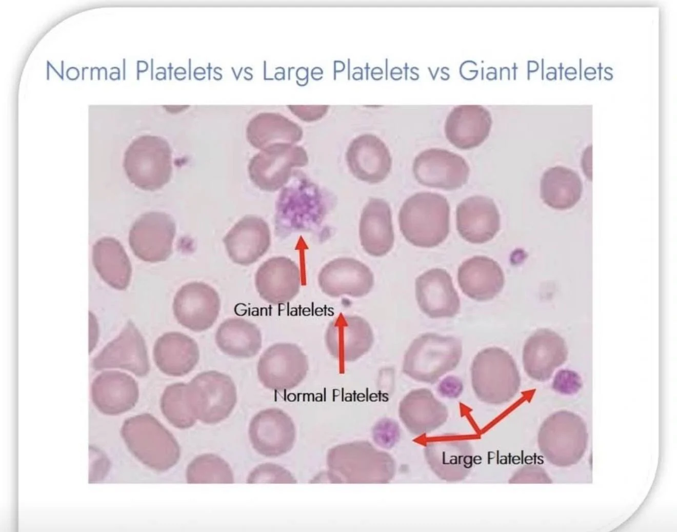
”Differences Between Normal Platelets, Large Platelets, and Giant Platelets
Platelets are essential components of blood that play a major role in blood clotting and hemostasis.
They can appear in various sizes and morphologic forms, as described below:
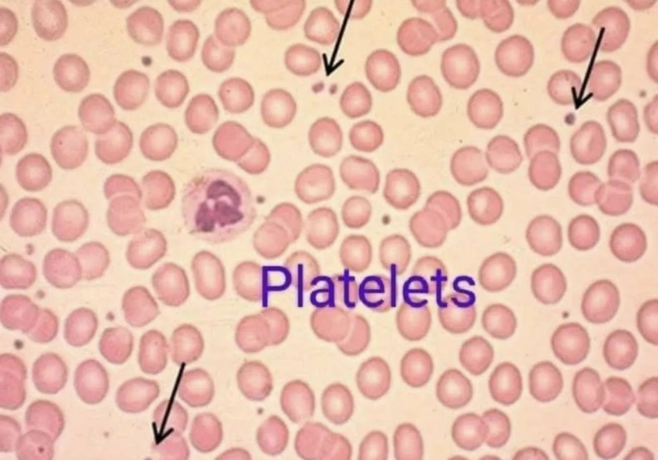
Normal Platelets
- Normal platelets are small, measuring approximately 2–3 micrometers in diameter.
- They are clearly smaller than red blood cells (RBCs).
- Morphologically, they are usually round to oval with evenly distributed purple granules within the cytoplasm.
- The presence of platelets with normal size and adequate numbers generally reflects balanced platelet production and normal bone marrow function.
Large Platelets
- Large platelets are bigger than normal platelets but are generally still smaller than red blood cells.
- They are commonly seen in conditions associated with increased platelet production, such as excessive platelet destruction in Immune Thrombocytopenia (ITP) or during recovery after blood loss.
- These platelets are often referred to as ‘young platelets’ because newly released platelets from the bone marrow tend to be larger and contain more prominent granules.
- Therefore, the presence of large platelets usually indicates an active and appropriate bone marrow response to platelet loss.
Giant Platelets
- Giant platelets are extremely large platelets that may approach or even exceed the size of red blood cells.
- They are considered clinically significant abnormalities and are often associated with inherited platelet disorders or abnormalities in megakaryocyte maturation.
Conditions associated with giant platelets include:
- Bernard–Soulier Syndrome
- May–Hegglin Anomaly
- Gray Platelet Syndrome
- Myeloproliferative disorders
- Severe platelet destruction or abnormal platelet production states
Laboratory Significance
The presence of large or giant platelets is highly important in hematology because automated hematology analyzers may mistakenly classify these oversized platelets as red blood cells. This can result in falsely low platelet counts or pseudothrombocytopenia.
Therefore, peripheral blood smear examination under the microscope remains essential for accurate platelet morphology assessment.
Summary
- Normal Platelets – Small, uniform in size, and clearly smaller than RBCs
- Large Platelets – Larger than normal, usually associated with increased platelet production
- Giant Platelets – Extremely large platelets, commonly associated with hematologic and inherited platelet disorders.”

Stay updated with Hemostasis Today.
-
Jun 30, 2026, 04:12Maia Meier: Driving Change for Women and Girls with Bleeding Disorders
-
Jun 30, 2026, 03:19Wolfgang Miesbach: Weight-Bearing Lunge Test for Ankle Assessment in Haemophilia?
-
Jun 30, 2026, 03:06Salvatore Brugaletta: Is Twelve Months of DAPT Still Too Long After ACS?
-
Jun 30, 2026, 02:55Edward Lee Carter: How A Landmark Stewardship Shift Reduced Unnecessary Antiplatelet Use Across VISN 8
-
Jun 30, 2026, 02:43Nicolas Gendron: More than a Research Fellowship – Leaving the Wagner Lab
-
Jun 30, 2026, 02:29Wolfgang Miesbach: Can We ‘Pre‑Tame’ the Immune System to Unlock Better AAV8 FVIII Expression?
-
Jun 30, 2026, 02:21Mohammad Khalil: Evaluating DAPT Duration after Percutaneous Coronary Intervention
-
Jun 30, 2026, 02:11Rishdha Roshad: Activated Clotting Time – Adult vs Pediatric
-
Jun 30, 2026, 02:02Julia Castillo González: Role of Cortistatin During the Acute and Subacute Phases of Ischemic Stroke