

Tushar Namdeo: Platelets in Different Diseases
Tushar Namdeo, Diagnostic Laboratory Specialist, shared a post on LinkedIn:
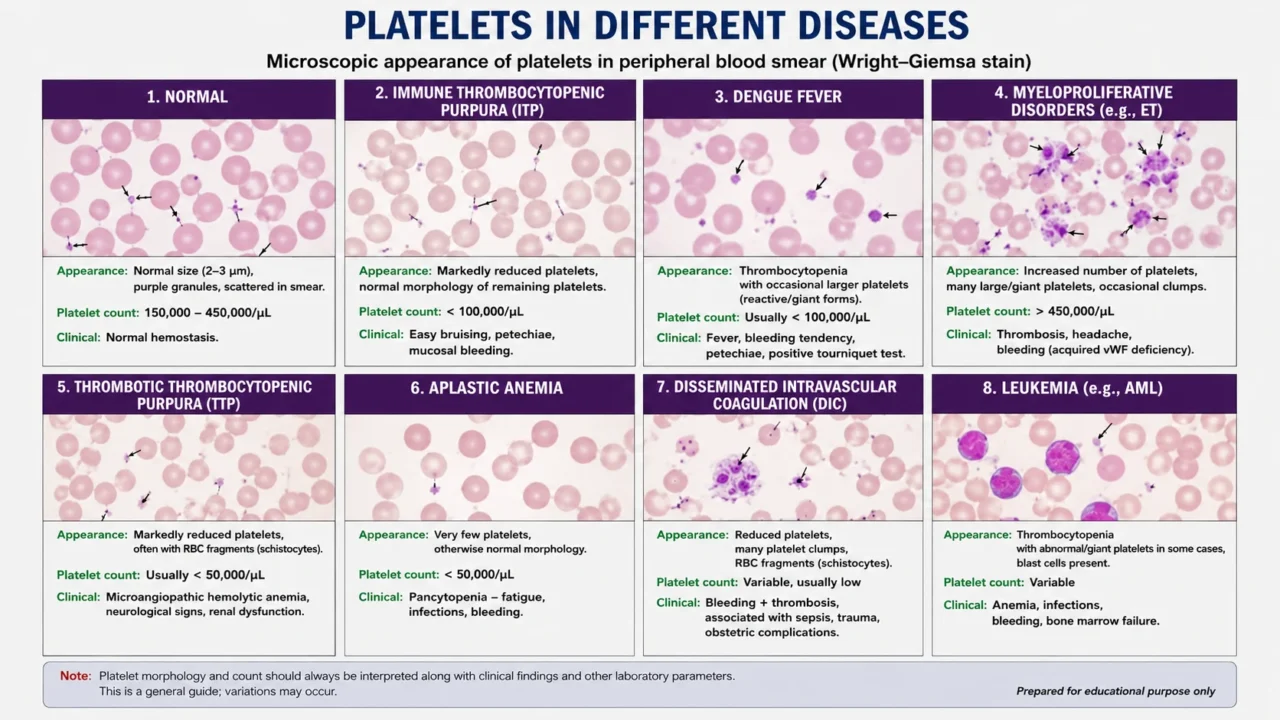
“This image is an educational microscopic guide to platelet appearance in different diseases using peripheral blood smear concepts.
Platelets in different conditions
1. Normal
- Platelets are small, purple-staining cell fragments.
- Normal platelet count: 150,000–450,000/µL.
- Usually scattered individually between RBCs.
2. ITP – Immune Thrombocytopenic Purpura
- Platelet count is typically low.
- Remaining platelets may include larger or young platelets.
- Clinical features can include petechiae, bruising and mucosal bleeding.
3. Dengue fever
- Thrombocytopenia is common.
- Platelet count can fall significantly during the illness.
- Larger or reactive platelets may sometimes be seen.
- Clinical interpretation should always consider the patient’s disease stage and other results.
4. Myeloproliferative disorders
- Platelet count may be markedly increased.
- Large or giant and variable platelet forms may be seen.
- Abnormal platelet function can occur despite a high count.
5. TTP – Thrombotic Thrombocytopenic Purpura
- Platelets are usually markedly decreased.
- Schistocytes (RBC fragments) may be present.
- This is an important hematological emergency requiring urgent clinical evaluation.
6. Aplastic anemia
- Platelet production is severely reduced because of bone-marrow failure.
- Very few platelets may be seen on the smear.
- Often associated with pancytopenia.
7. DIC – Disseminated Intravascular Coagulation
- Platelets are commonly reduced because they are consumed.
- Schistocytes and abnormal clotting may be present.
- Can occur with severe infection/sepsis, trauma and other serious conditions.
8. Leukemia
- Platelet count may be low because of bone-marrow involvement.
- Platelet morphology can be abnormal.
- Abnormal blast cells may be visible depending on the type of leukemia.
Important laboratory point
Never interpret platelet morphology alone. Correlate the peripheral smear with CBC or platelet count, MPV, clinical findings, coagulation tests and other relevant laboratory results. A smear finding by itself does not establish a diagnosis.”
Stay updated with Hemostasis Today.
-
Oct 16, 2026, 13:10Emanuel Sueldo: CAHT 2026 Celebrates Scientific Progress and Team Achievements
-
Oct 16, 2026, 13:05Maha Othman: Queen’s Health Sciences Advances CAT Risk Models and Pediatric ISTH-BAT Cut-Offs
-
Oct 16, 2026, 13:03Thrombosis Awareness Must Lead to Timely Action – Roche
-
Oct 16, 2026, 12:49Wolfgang Miesbach: 5-Year CBDR Data Confirms Emicizumab Safety and Efficacy in Hemophilia A
-
Oct 16, 2026, 12:40Edward Lee Carter: How Factor XI Inhibition Could Transform Anticoagulation Therapy
-
Oct 16, 2026, 12:40Marc Carrier: Looking Ahead to ISTH 2027 in Vancouver
-
Oct 16, 2026, 12:30Bob Lempke: How One Blood Donation Can Save Up to Three Lives
-
Oct 16, 2026, 12:30Why VWF Therapies Belong on the EU Critical Medicines List – EHC
-
Oct 16, 2026, 12:17Exploring Endothelial Cells, Genetic Fate Mapping, and Revascularization – ISTH