

A CT-Based Clinical Overview from Muhammad Abubakar: Stroke and Intracranial Hemorrhages
Muhammad Abubakar, Member of Congress of Neurological Surgeons (CNS), shared on LinkedIn:
”Stroke and Intracranial Hemorrhages — A CT-Based clinical Overview
Stroke is a radiological and clinical emergency.
Rapid CT interpretation can be lifesaving.
Here’s a structured breakdown every medical student and clinician should master:
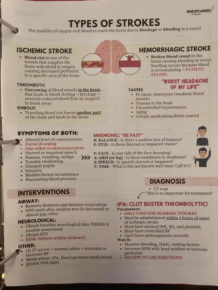
STROKE — THE BIG PICTURE
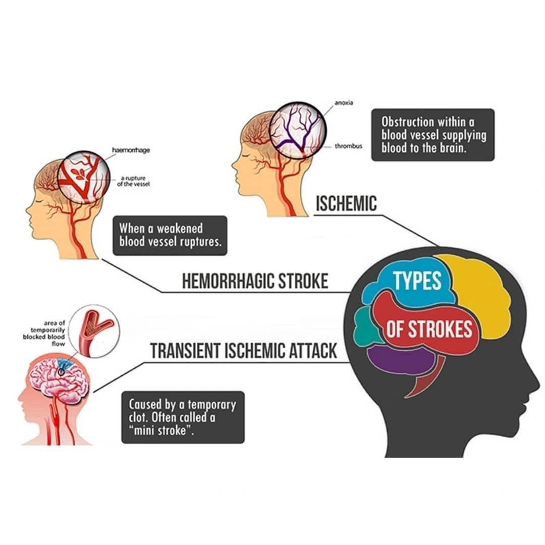
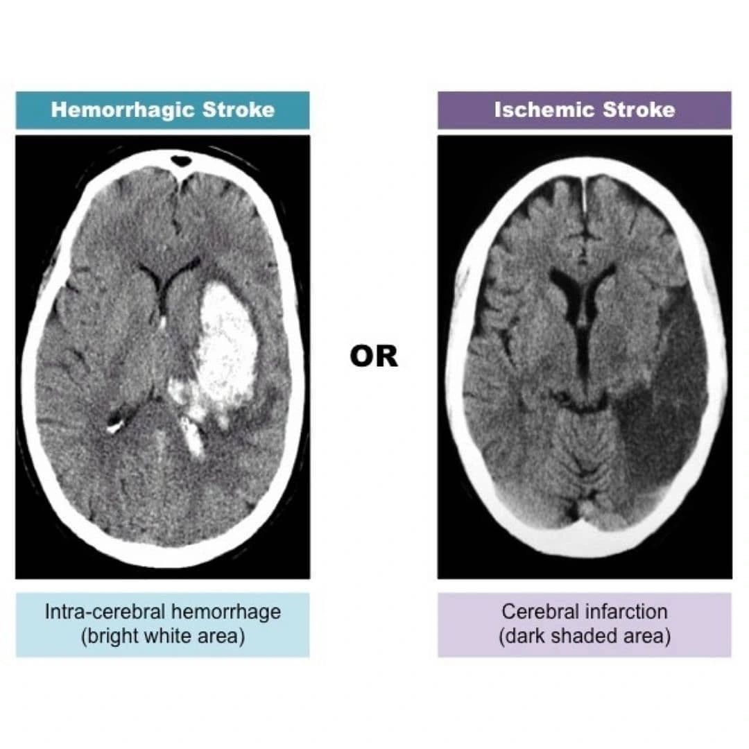
Ischemic Stroke (≈85%)
Cause: Arterial occlusion → cerebral infarction
Early CT (may be normal):
• Loss of gray–white matter differentiation
• Insular ribbon sign
• Hyperdense MCA sign
Late CT:
• Hypodense wedge-shaped infarct
• Sulcal effacement
• Mass effect due to edema
Rule: Always exclude hemorrhage on CT before thrombolysis.
Hemorrhagic Stroke (≈15%)
Cause: Vessel rupture → bleeding
CT hallmark:
• Hyperdense (white) blood
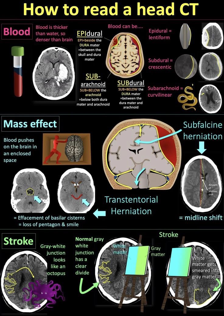
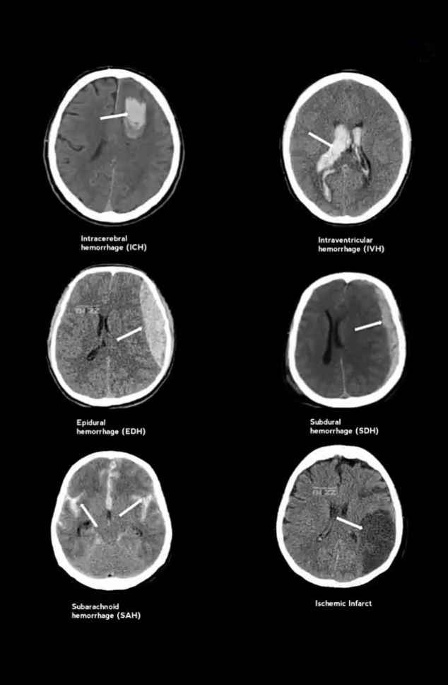
EXTRA-AXIAL HEMORRHAGES
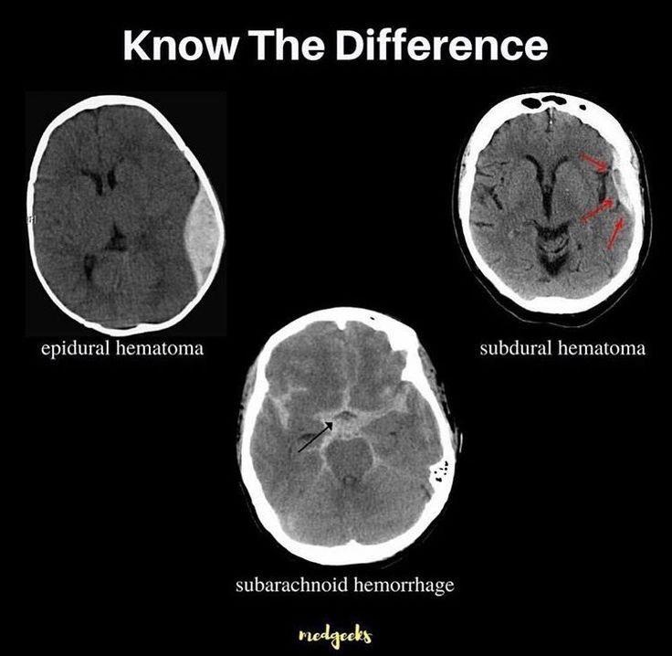
Epidural Hemorrhage (EDH)
Cause: Middle meningeal artery injury (trauma)
CT Findings:
- Biconvex / lentiform shape
- Does not cross suture lines
- Classically associated with lucid interval
Subdural Hemorrhage (SDH)
Cause: Bridging vein tear
CT Findings:
- Crescent-shaped collection
- Crosses suture lines (not midline)
Acute → white – hyperdense
Chronic → black – hypodense
Subarachnoid Hemorrhage (SAH)
Cause: Berry aneurysm rupture / trauma
CT Findings:
- Hyperdensity in sulci, cisterns, fissures
- “Star-shaped” basal cisterns
- Sudden worst headache of life
INTRA-AXIAL HEMORRHAGES
Intracerebral / Intraparenchymal Hemorrhage (ICH)
Cause: Hypertension, cerebral amyloid angiopathy
CT Findings:
- Localized hyperdense bleed within brain parenchyma
- Surrounding hypodense edema
- Common sites: basal ganglia, thalamus, pons, cerebellum
Intraventricular Hemorrhage (IVH)
Cause: Extension of ICH / neonatal germinal matrix bleed
CT Findings:
- Hyperdense blood layering in ventricles
- Ventricular dilatation → hydrocephalus
ONE-LINE MEMORY AID
CT Rules to Remember:
Blood = White
Ischemia = Dark (late)
Extra-axial → shape matters
Intra-axial → location matters
Mastering CT patterns equals faster diagnosis plus better neurological outcomes”

Stay updated with Hemostasis Today.
-
Jul 2, 2026, 17:05Haroun Gajraj: Understanding the Technology Behind Spider Vein Treatment
-
Jul 2, 2026, 16:44Ney Carter Borges: Is It Time to Personalize Antiplatelet Therapy?
-
Jul 2, 2026, 16:22Introducing a New Resource for the ITP Community – ITP Support Association
-
Jul 2, 2026, 16:22Shinto Francis Thekkudan: Delegates Visiting our BMT and Hematology Department from Kazhakstan
-
Jul 2, 2026, 16:13William Aird: When Deciding whether an Adult Patient is Anemic, Which Do You Usually Use?
-
Jul 2, 2026, 16:08Supporting Bleeding Disorders Community and Celebrating ASPEH Milestone – WFH
-
Jul 2, 2026, 15:59Raul Santos: Why Does LDL-C Remain Elevated in Many Patients?
-
Jul 2, 2026, 15:16Tim Patel: How Regular Movement Helps Keep Blood Flowing Throughout the Day
-
Jul 2, 2026, 13:16Nathan Connell: Is It Time to Rethink What We Tell Hemophilia B Carriers About Pregnancy?