

Gurpreet Kaur Sagoo: The Power of AI Based Morphological Fingerprinting in Predicting Myelofibrosis Progression
Gurpreet Kaur Sagoo, Professor at Armed Forces Medical College, shared a post on LinkedIn about a recent article she and her colleagues co-authored, published in Indian Journal of Hematology and Blood Transfusion, adding:
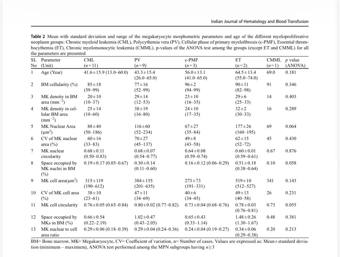
“Morphometric analysis of megakaryocytes plays a central role in diagnosing and subclassifying Philadelphia-chromosome negative Myeloproliferative Neoplasms (MPNs), as specific cellular and spatial abnormalities distinguish Essential Thrombocythemia (ET), Polycythemia Vera (PV), and Primary Myelofibrosis (PMF).
While manual histopathological evaluation is a standard World Health Organization (WHO) requirement, digital morphometry and machine learning workflows on bone marrow trephine biopsies provide precise, reproducible, and automated quantification of these parameters to avoid subjective variation.
Primary Morphometric Parameters Analyzed Digital image analysis systems typically measure several core quantitative indicators from H&E or immunohistochemically stained sections:
- Cellular Size and Area: Calculated via the cytoplasmic major diameter and total cytoplasmic area.
- Nuclear Area and Perimeter: Measures the total footprint of the nucleus and its boundary length.
- Nuclear-to-Cytoplasmic (N:C) Ratio: Derived by dividing the nuclear area by the overall cytoplasmic area.
- Nuclear Shape Indices: Quantified via the Nuclear Roundness Factor and Nuclear Contour Ratio to capture the irregularity or lobation profile.
- Topographical Clustering and Density: Quantitative spatial alignment mapping whether cells form loose or tight clusters (3 or more juxtaposed cells) and their spatial distance from bone trabeculae or blood sinuses.
- Advanced and Predictive Morphometric markers:
Megakaryocytic Activation (M-ACT Index):
A specialized morphometric index scored on the coexistence of megakaryocytic emperipolesis (intact neutrophils passing through megakaryocyte cytoplasm), tight cluster count, and dense collagen fiber perimeters.
High M-ACT indices mathematically correlate with higher JAK2 V617F allele burden and predict rapid progression to secondary myelofibrosis.
AI-Based Morphological Fingerprinting: Advanced neural networks extract up to 9 distinct megakaryocytic phenotypes from bone marrow images based on shape variations, generating cohort-wide radar plots.
This high-throughput tool isolates true MPNs from reactive mimics (like immune thrombocytopenia or reactive thrombocytosis) with an area under the curve (AUC) accuracy of up to 0.95.”
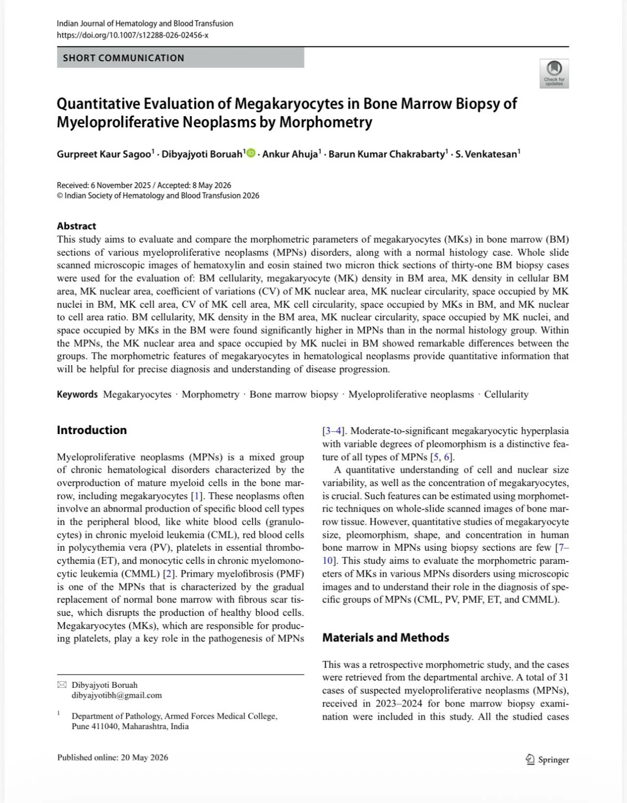
Title: Quantitative Evaluation of Megakaryocytes in Bone Marrow Biopsy of Myeloproliferative Neoplasms by Morphometry
Authors: Gurpreet Kaur Sagoo, Dibyajyoti Boruah, Ankur Ahuja, Barun Kumar Chakrabarty, S. Venkatesan

Stay updated on all scientific advances with Hemostasis Today.
-
Jul 13, 2026, 11:25Luisa Müller: Advancing Anticoagulation Strategies for VITT at ISTH 2026
-
Jul 13, 2026, 10:49Joshua Zeidner: Advancing Menin Inhibitor Therapy in MD Education AML-ALL US Focus 2026
-
Jul 13, 2026, 10:25Flora Peyvandi: A Practical Approach to ALT Elevation After Gene Therapy at ISTH 2026
-
Jul 13, 2026, 10:02Jessica Garcia: Where Science and Community Come Together at ISTH 2026
-
Jul 13, 2026, 09:46Fabrice Cognasse: Is There Such a Thing as the Perfect Platelet Product?
-
Jul 13, 2026, 09:42Cancer-Associated Arterial Thromboembolism: Redefining the Thrombotic Burden in Oncology
-
Jul 13, 2026, 05:59Connecting and Collaborating at ISTH 2026 – NNHF
-
Jul 13, 2026, 05:58Lara Monica: Back at ISTH – Looking Forward to Science and Reconnections!
-
Jul 13, 2026, 05:57Barbara Adams Krolak: Exciting Updates From the ISTH 2026 Regulatory Session