Jan Sloves: When Stent Placement Misses the Mark – A Lesson in Precision and Follow-Up
Jan Sloves, President and Consultant at Vascular Imaging Professionals LLC, shared on LinkedIn:
”When Stent Placement Misses the Mark: A Lesson in Precision and Follow-Up
This is a great real-world example of why precise access and follow-up imaging matter.
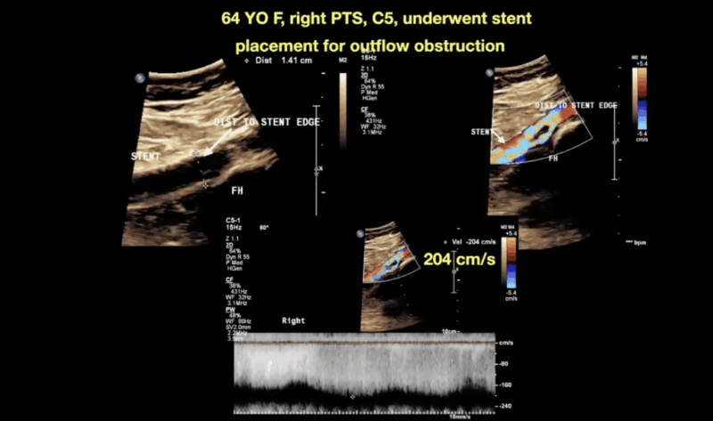
Here, we’re looking at a 64-year-old female with right-sided post-thrombotic changes (C5) and a healed venous ulcer.
She previously underwent stent placement for significant outflow obstruction — but the access site was positioned too high.
As a result, the true lesion was missed, leaving behind venous fibrosis and aliasing consistent with a high-grade venous stenosis (~200 cm/s).
Despite intervention, the patient remained symptomatic and ultimately required revision in the cath lab.
In this case, I’m demonstrating how to assess the diameter and velocity ratio — greater than 5 — which meets diagnostic criteria for left renal vein compression.
Technical pearls:
Optimize grayscale to clearly delineate anatomy.
Use calipers to measure both the maximally obstructed and dilated segments.
Advance your Doppler sample through the narrowest point to capture the peak velocity for an accurate ratio.
It’s straightforward — but small details like these define accuracy, guide treatment, and ultimately improve patient outcomes.
Want to see more practical vascular cases and scanning strategies like this?
Join our growing education hub at Ultrasound Unlocked.
— where physicians and sonographers share real cases, refine technique, and grow together.”

Stay updated with Hemostasis Today.
-
Feb 23, 2026, 11:37Charles Okyere Boadu: Blood Donation Helps Lower Your Risk of Stroke and Organ Damage
-
Feb 23, 2026, 11:29Emma Lefrancais: Uncovering A Key Role for The IL-33/ST2 Axis in Platelet Biology with Lucie Gelon
-
Feb 22, 2026, 14:16Ilenia Calcaterra: From Representation to Intellectual Independence in Women in Science
-
Feb 22, 2026, 13:27Pete Stibbs: New AHA and ACC PE Guidelines Finally Align with Real Clinical Practice
-
Feb 22, 2026, 10:39Tagreed Alkaltham: Fibrinogen Concentrate Is a Deliberate Clinical Choice in Acute Bleeding
-
Feb 22, 2026, 09:38Abdulrahman Nasiri: Significant Shifts In The 2026 AHA/ACC Guidelines for Acute Pulmonary Embolism
-
Feb 22, 2026, 09:22Shiny K. Kajal: Not All Transfusion Reactions Are Immunohematologic Incompatibilities
-
Feb 22, 2026, 09:12Arun V J։ The Hidden Risks in Every Blood Bag
-
Feb 22, 2026, 08:56Parandzem Khachatryan։ How Hard Is It to Be a Mom, a Wife, a Professor, and a Doctor All at Once?

