


Fredrick Chalinga: Negative DAT Does Not Mean No Hemolysis
Fredrick Chalinga, Medical Technologist at Aga Khan University Hospital, shared a post on LinkedIn:
”The patient is clearly hemolyzing… but the DAT is negative?
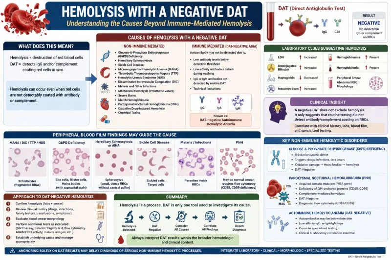
One of the most confusing situations in transfusion medicine and hematology is encountering evidence of hemolysis with a negative Direct Antiglobulin Test (DAT).
Many clinicians assume:
Positive hemolysis means positive DAT.
But in reality, not all hemolytic anemia is immune-mediated, and even immune hemolysis can occasionally produce a negative DAT. This is where laboratory correlation becomes critical.
What does this mean?
Hemolysis indicates destruction of red blood cells, which may be immune-mediated, mechanical, infectious, chemical, hereditary, or microangiopathic.
The DAT specifically detects IgG and/or complement coating red cells in vivo.
Therefore, hemolysis can occur even when red cells are not detectably coated with antibody or complement.
Common causes of hemolysis with a negative DAT include:
- G6PD deficiency
- Hereditary spherocytosis
- Sickle cell disease
- Microangiopathic hemolytic anemia (MAHA)
- TTP, HUS, and DIC
- Malaria and other infections
- Mechanical hemolysis from prosthetic valves
- Severe burns
- March hemoglobinuria
- Paroxysmal nocturnal hemoglobinuria (PNH)
- Oxidative drug-induced hemolysis
Important to note:
Some cases of autoimmune hemolytic anemia (AIHA) may also appear DAT-negative.
Why?
Because antibody levels may be below the detection threshold, low-affinity antibodies may detach during washing, IgA or IgM antibodies may not be detected by routine DAT reagents, and technical limitations can occur.
This is known as DAT-negative autoimmune hemolytic anemia.
Laboratory clues suggesting hemolysis include:
- Increased LDH
- Increased unconjugated bilirubin
- Reduced haptoglobin
- Reticulocytosis
- Hemoglobinemia or hemoglobinuria
- Spherocytes or schistocytes on peripheral smear
Peripheral blood film findings may guide the cause:
- Schistocytes may suggest MAHA, DIC, or TTP.
- Bite cells and blister cells may suggest G6PD deficiency.
- Spherocytes may suggest hereditary spherocytosis or AIHA.
- Sickled cells may suggest sickle cell disease.
- Parasites may suggest malaria.
Clinical insight:
A negative DAT does not exclude hemolysis. It only suggests that routine testing did not detect antibody or complement coating on red blood cells.
That distinction is extremely important. In transfusion medicine, anchoring solely on DAT results may delay diagnosis of serious non-immune hemolytic processes.
The blood film, hemolysis markers, clinical history, and specialized testing remain essential.
Key takeaway:
Hemolysis is a process. DAT is only one tool used to investigate its cause. Always interpret DAT results within the broader hematologic and clinical context.
Have you encountered challenging DAT-negative hemolysis cases in practice?”
Stay updated with Hemostasis Today.
-
Oct 20, 2026, 04:13Ifeanyichukwu Ifechidere: DOACs Are Not the Answer to Every Anticoagulation Question
-
Oct 20, 2026, 04:04Sadia Kamal: Honored to Receive the Outstanding Abstract Award at the 20th Midwest Platelet Conference
-
Oct 20, 2026, 04:03Yu-Hsuan Chen: One Antibody, Two Diseases from FcRn Biology to Clinical Translation
-
Oct 20, 2026, 03:49Domenico Simone Castiello: Rethinking Prolonged DAPT in Multivessel PCI – Insights from the DAPT-MVD Trial
-
Oct 20, 2026, 03:37Rucha Patil: Scaling POC Diagnostics for Haemophilia A and VWD Across India
-
Oct 20, 2026, 03:31Zena Vexler: Targeting Neutrophil Elastase and NETosis to Mitigate Infection-Triggered Childhood Stroke
-
Oct 20, 2026, 03:24Chaza Alaji: Diagnosing Protein C and Protein S Deficiency
-
Oct 20, 2026, 03:24Mou Nandi: How Maternal Health During Pregnancy May Shape Infant Development
-
Oct 20, 2026, 03:13Sumyea Kabir: Pre-Analytical Quality in Phlebotomy – Preventing In Vitro Hemolysis