


Ayuka King-Percival: How Lupus Anticoagulant Testing Works
Ayuka King-Percival, Senior Medical Technologist, Head of Hematology at LABORATORY, Adjunct Lecturer at College of Science, Technology and Applied Arts of Trinidad and Tobago-COSTAATT, shared a post on LinkedIn:
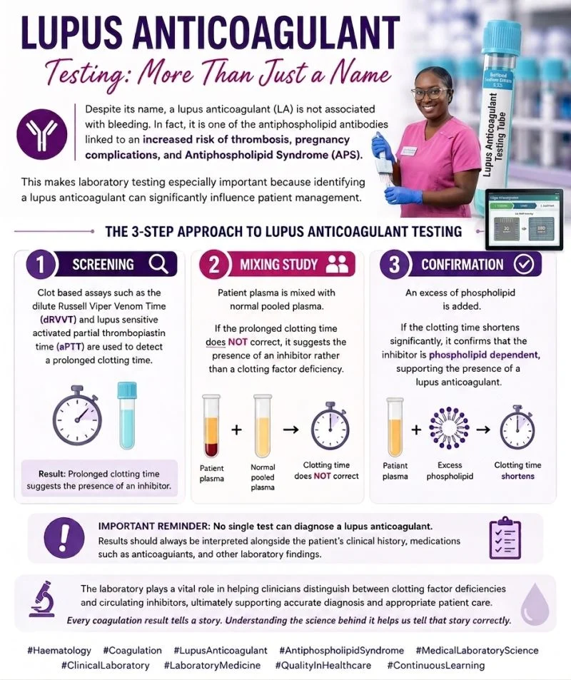
“Lupus Anticoagulant Testing: More Than Just a Name.
Despite its name, a lupus anticoagulant (LA) is not associated with bleeding.
It is an antiphospholipid antibody detected by its effect on phospholipid dependent coagulation assays and is associated with conditions such as thrombosis, pregnancy complications, and Antiphospholipid Syndrome (APS).
Laboratory investigation involves three key stages:
1. Screening
Tests such as dilute Russell Viper Venom Time (dRVVT) and lupus sensitive activated partial thromboplastin time (aPTT) identify prolonged clotting times.
2. Mixing Study
Patient plasma is mixed with normal pooled plasma. Failure to correct suggests the presence of an inhibitor rather than a clotting factor deficiency.
3. Confirmation
Excess phospholipid is added. Shortening of the clotting time demonstrates phospholipid dependence, supporting the presence of a lupus anticoagulant.
A laboratory teaching analogy: Understanding the confirmation step
Imagine a car park with 20 available spaces.
The lupus anticoagulant is like 15 cars occupying those spaces, leaving limited room for phospholipid dependent coagulation reactions.
With reduced available phospholipid in the assay, the lupus anticoagulant can exert its inhibitory effect, resulting in a prolonged clotting time during screening.
Now imagine expanding the car park to 200 spaces.
The same 15 cars remain, but there are now 185 spaces available.
The additional phospholipid helps reduce the inhibitory effect of the lupus anticoagulant, allowing the coagulation reaction to proceed more normally and shortening the clotting time.
This is a simplified analogy.
Lupus anticoagulants do not literally occupy phospholipid molecules.
Instead, they interfere with phospholipid dependent coagulation reactions, which is why excess phospholipid can reduce their effect within the assay.
A lupus anticoagulant result should always be interpreted alongside the patient’s clinical history, anticoagulant therapy, potential assay interference, and other antiphospholipid antibody results.
The laboratory plays a vital role in translating complex coagulation findings into meaningful clinical information.
Every coagulation result tells a story.
Understanding the science behind it helps us tell that story correctly.”
Stay updated with Hemostasis Today.
-
Sep 20, 2026, 13:29Factor XI Deficiency and the Challenge of Unpredictable Bleeding – EHC
-
Sep 20, 2026, 13:18Cindy Howry: FDA Approves First Treatment for Ataxia-Telangiectasia
-
Sep 20, 2026, 13:08Mohammad Salar Amoli: When Routine Coagulation Tests Miss von Willebrand Disease
-
Sep 20, 2026, 12:55Stelios Andreadis։ Human Salivary Gland Organoids Offer New Potential for Regenerative Medicine
-
Sep 20, 2026, 12:43Obi Light Ogbonnia: The Power of Lived Experience in Sickle Cell Care
-
Sep 20, 2026, 12:34Sabrine Mekni: Advancing HSCT for Children With Fanconi Anemia
-
Sep 20, 2026, 12:11Giacomo Buso: Silent Atherosclerosis Begins Earlier Than Expected
-
Sep 20, 2026, 12:0310 Posts Not to Miss This Week
-
Sep 20, 2026, 11:27Edward Lee Carter: Prasugrel Dosing Matters in P2Y12 Inhibitor Comparisons After PCI