
Jan Sloves: Turning Routine Duplex into a Multiparametric Evaluation of Limb Edema
Jan Sloves, President and Consultant at Vascular Imaging Professionals LLC, shared a post on LinkedIn about a recent article by Luca Palombi et al, published in Phlebology:
“We Often Use Venous Duplex to ‘exclude DVT and Reflux’ in Patients with Limb Swelling-But How Often Do We Deliberately Look for Lymphedema?
A just published pictorial essay by Luca Palombi et al. in Phlebology 2026 highlights how duplex ultrasound can directly visualize lymphatic‑related tissue changes and help stage lymphedema, not just rule out venous disease.
The authors remind us that lymphedema is still frequently misdiagnosed as chronic venous insufficiency or lipedema despite distinct tissue changes on ultrasound.
Key sonographic insights that stood out to me:
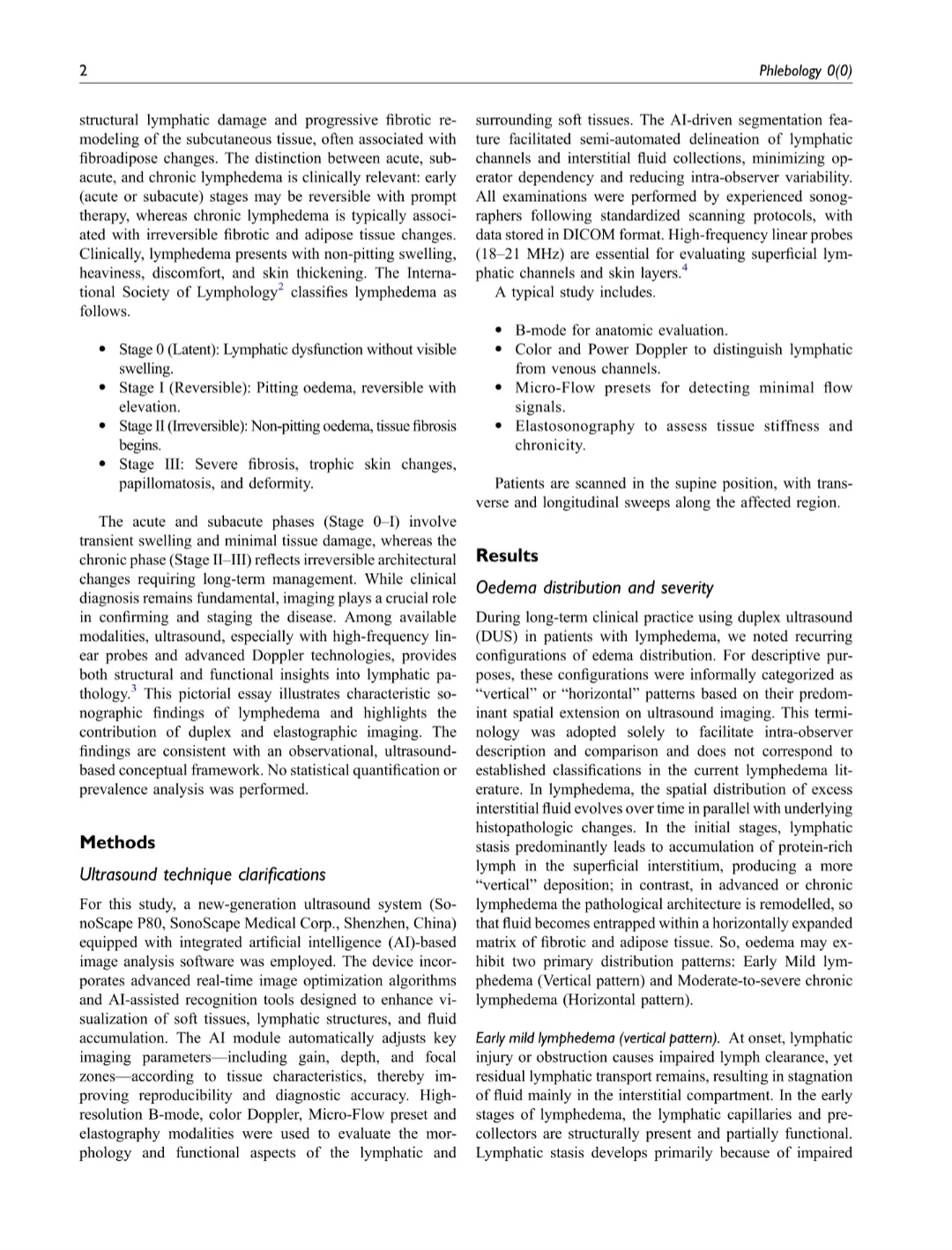
- Early/mild lymphedema tends to show a ‘vertical‘ pattern: fluid tracking from the dermis toward the superficial fascia along perivascular and peri-lymphatic planes
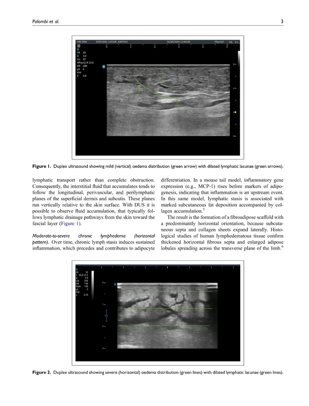
- Moderate–severe chronic lymphedema shows a ‘horizontal‘ pattern: transversely expanded, fibroadipose subcutaneous tissue with hypoechoic lymphatic lacunae
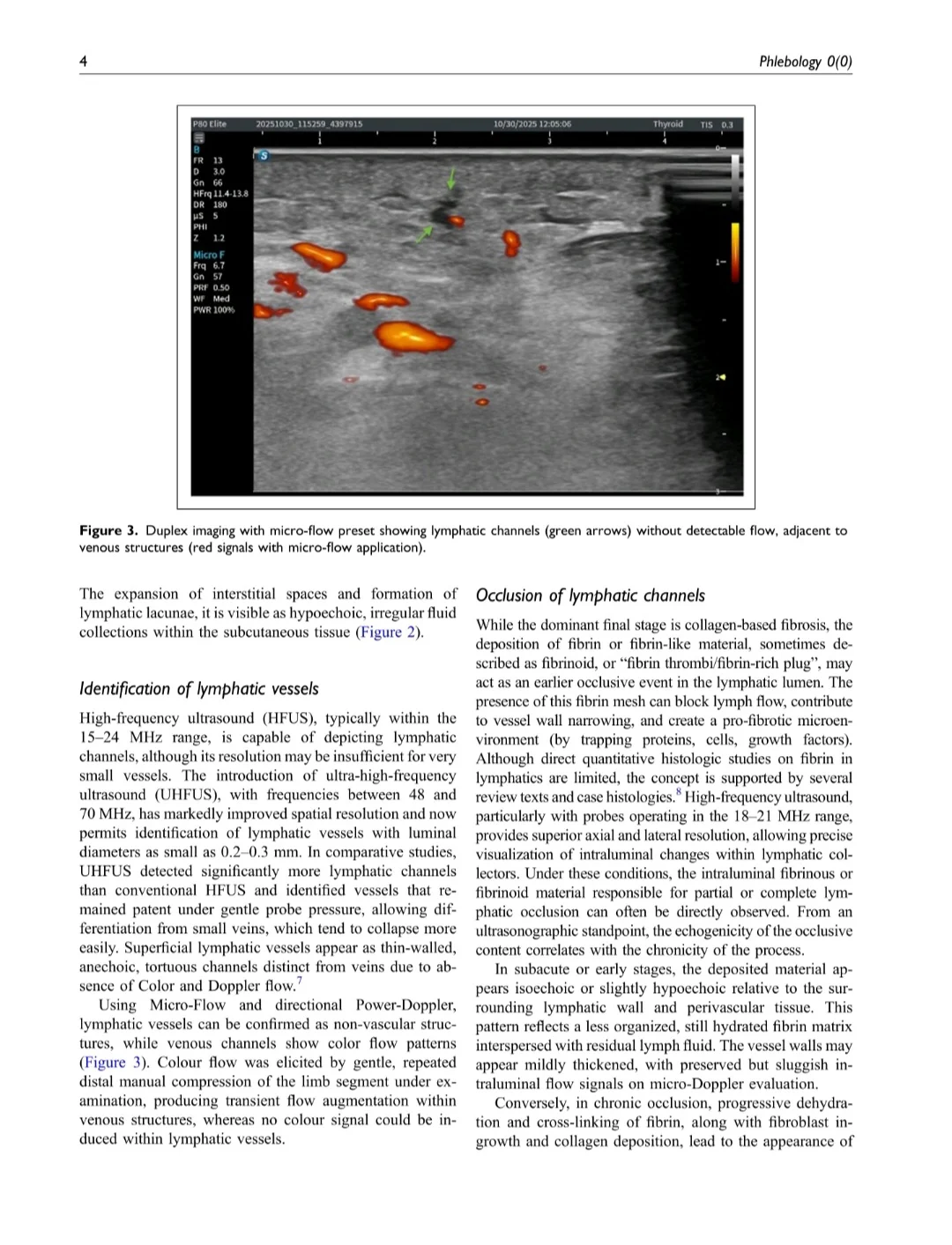
- Superficial lymphatics appear as thin‑walled, tortuous, non‑compressible, Doppler‑silent channels, even with distal manual compression, helping distinguish them from small veins
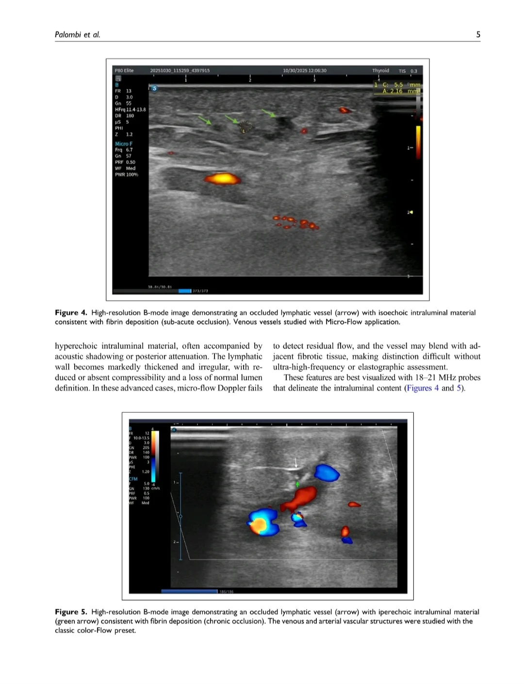
- High‑frequency imaging and micro‑flow presets can show intraluminal fibrinous plugs in chronically obstructed lymphatics, offering a window into irreversible damage
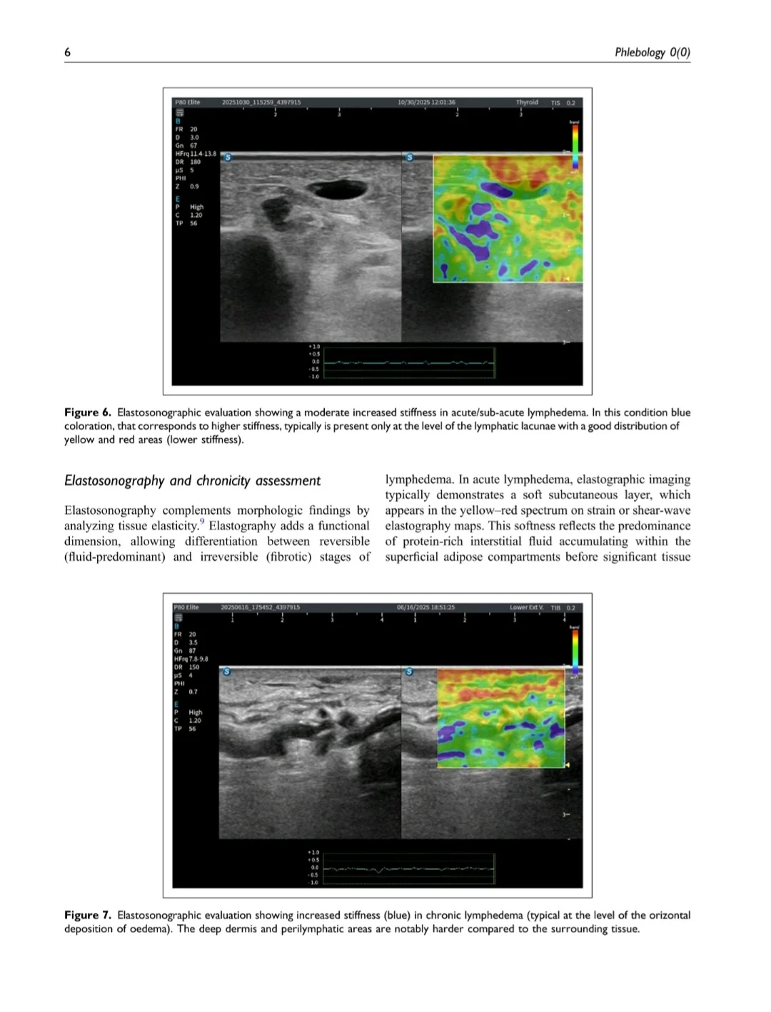
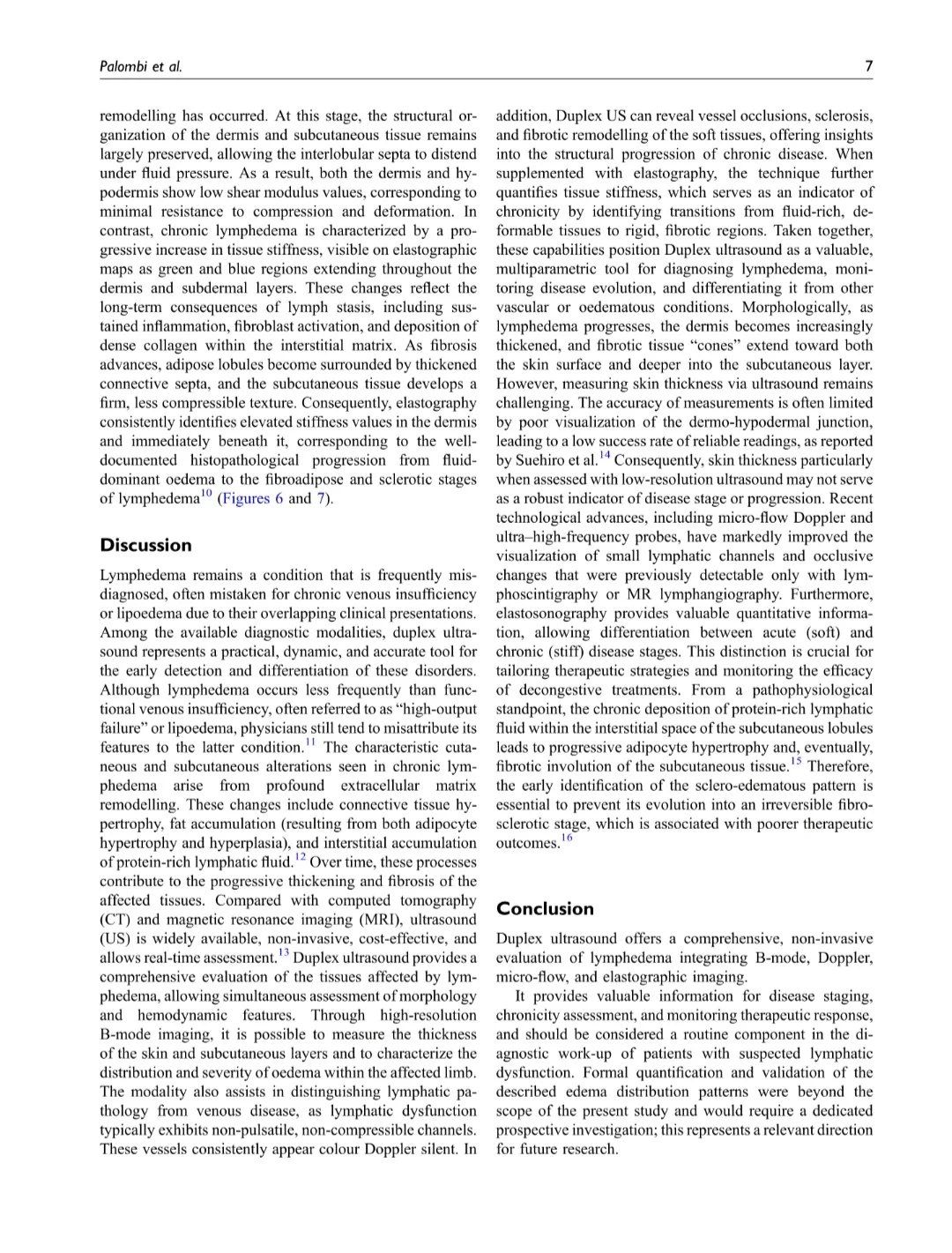
- Elastography adds a functional layer: ‘soft‘ fluid‑predominant acute disease versus ‘stiff’ fibrotic chronic disease, supporting assessment of chronicity and treatment planning
- The examinations used high‑frequency probes plus micro‑flow and elastography with AI‑assisted image optimization to enhance lymphatic and soft‑tissue visualization and reduce operator dependency. The authors utilized transducer frequencies from 18-21 MHz for superficial lymphatic channels and skin layers and ultra-high frequencies 48-70 MHz for depicting even smaller lymphatic vessels
For those scanning ‘just to rule out venous disease’, this paper is a useful reminder that a focused lymphatic/soft‑tissue assessment can turn a routine duplex into a genuinely multiparametric evaluation of limb edema and should be part of the standard work‑up when lymphedema is on the table.
Subscribe to the only newsletter built for vascular pros.”
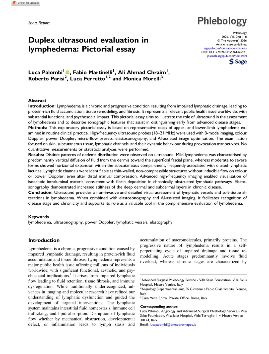
Title: Duplex ultrasound evaluation in lymphedema: Pictorial essay
Authors: Luca Palombi, Fabio Martinelli, Ali Ahmad Chraim, Roberto Parisi, Luca Ferretto, Monica Morelli
Read the Full Article on Phlebology

Other articles featuring Jan Sloves on Hemostasis Today.
-
Jul 11, 2026, 08:50Heghine Khachatryan: Multidisciplinary PERTs Transform Pulmonary Embolism Care
-
Jul 11, 2026, 08:37Alejandro González Veliz: Key Takeaways from the 2026 ACC Scientific Statement on Antiplatelet Therapy
-
Jul 11, 2026, 08:35Rono Chonginio: The Power of Visual Storytelling in Healthcare and Non-Profits
-
Jul 11, 2026, 08:35Mavis Agnes Kisakye: Why Patient Identification Begins with an Informed Community
-
Jul 11, 2026, 08:35Leslie Lake: Compression Therapy in Venous Thromboembolism
-
Jul 10, 2026, 22:43Marignima Bennett: Grady Health System Earns the Highest Recognition for Excellence in Stroke Care
-
Jul 10, 2026, 21:31Robert Hamilton: Join NeuraSignal’s Webinar on Real-World Implementation of Robotic TCD in Stroke Care
-
Jul 10, 2026, 20:47New Machine Learning Models Improve Prediction of Thrombosis and Bleeding in Cancer – JTH
-
Jul 10, 2026, 20:39Evangelia Petsalaki: New Systems Biology Approach Identifies Biomarkers in Thrombotic APS