
Krishnakant Prasad/LinkedIn
May 9, 2026, 10:25
Krishnakant Prasad: A Dangerous Blood Clot That Can Lead to Pulmonary Embolism
Krishnakant Prasad, Associate Professor of Mathematics at Delhi Skill and Entrepreneurship University, shared a post on LinkedIn:
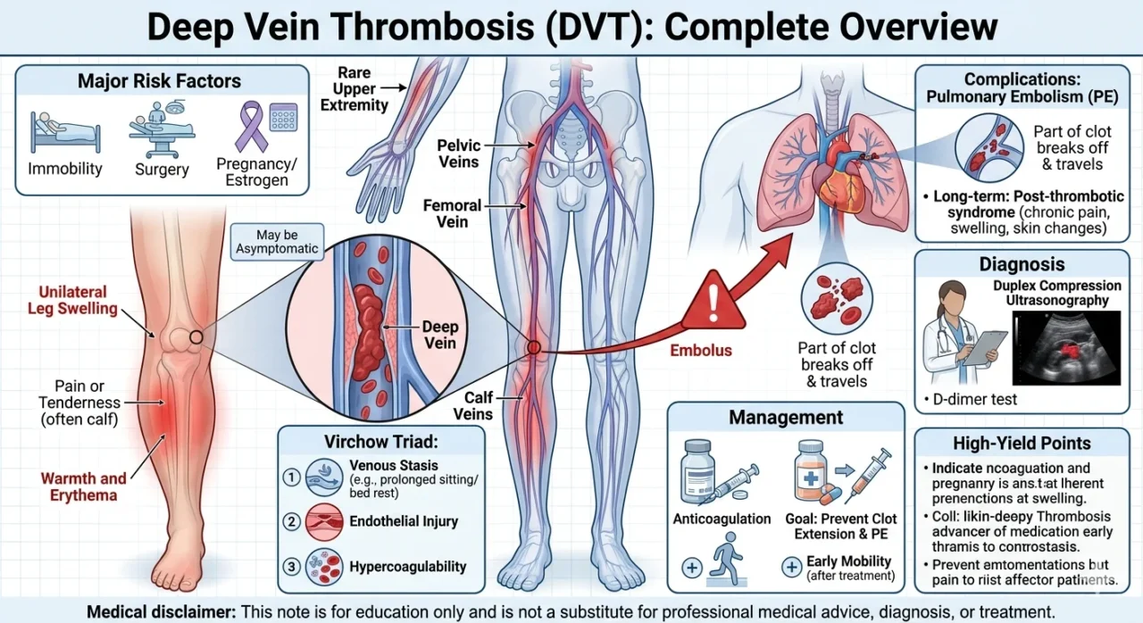
“Deep Vein Thrombosis (DVT):
Formation of a blood clot in a deep vein, usually in the leg or pelvis.
It is dangerous because part of the clot can break off and cause pulmonary embolism (PE).
Common Sites:
- Most often affects the calf, popliteal, femoral, or pelvic veins
- Less commonly can involve the upper extremity
Major Risk Factors:
- Immobility or prolonged bed rest
- Recent surgery, trauma, or hospitalization
- Cancer
- Pregnancy/postpartum, estrogen therapy, OCP use
- Prior DVT/PE or inherited/acquired thrombophilia
Pathophysiology:
- Classically related to Virchow triad
- Venous stasis
- Endothelial injury
- Hypercoagulability
Clinical Features:
- Unilateral leg swelling
- Leg pain or tenderness, often calf pain
- Warmth and erythema of the affected limb
- May have a feeling of heaviness or tightness
- Some patients are asymptomatic
Important Exam Clues:
- Usually one leg is more swollen than the other
- Calf tenderness may be present
- Clinical diagnosis alone is unreliable because signs are nonspecific
Complications:
- Pulmonary embolism is the most important acute complication
- Long-term complication: post-thrombotic syndrome with chronic pain, swelling, skin changes, or venous insufficiency
- Recurrent DVT can occur
Diagnosis:
- Duplex compression ultrasonography is the main test
- D-dimer may help rule out DVT in selected low-risk patients
- Clinical probability tools are often used before testing
Management:
- Anticoagulation is the main treatment
- Goal is to prevent clot extension and PE
- Some selected severe cases may need thrombolysis, thrombectomy, or filter placement
- Early mobility is generally allowed once treated
High-Yield Points:
- DVT is unilateral leg swelling plus pain plus warmth
- Biggest danger is pulmonary embolism
- Think of Virchow triad
- Ultrasound is the key diagnostic test
- Anticoagulation is the standard treatment
Medical disclaimer:
This note is for education only and is not a substitute for professional medical advice, diagnosis, or treatment.”

Stay updated with Hemostasis Today.
-
Jun 26, 2026, 18:28Daniel Pereira Monteiro: Why Is Beta Thalassaemia Major so Severe?
-
Jun 26, 2026, 18:27Pradip Pawar: Cell and Gene Therapy – Transforming Medicine in the 21st Century
-
Jun 26, 2026, 18:26Aviva Schwartz: How Can We Better Protect Patients After an Ischemic Stroke?
-
Jun 26, 2026, 18:25Does Reversing Factor Xa Inhibitors Really Carry as Much Thrombotic Risk as We Think? – RPTH Journal
-
Jun 26, 2026, 18:25Akshat Jain: Novel Therapies in Childhood Blood and Cancer Disorders
-
Jun 26, 2026, 16:57Abdul Muqtadir Abbasi: Anticoagulation in DVT Patients With Hidden Varices Requires Extreme Caution
-
Jun 26, 2026, 16:42Anesa Mulabecirovic: Why Von Willebrand Disease Takes Years to Diagnose in Women
-
Jun 26, 2026, 16:32Kaitlin Lichty: A New Identity Reflecting a Shared Legacy of Bleeding Disorders
-
Jun 26, 2026, 16:07Toong Youttananukorn: The WFH Registry Driving Better Care and Advocacy