


Abdul Mannan: Pencil Cells in IDA – Why Most, but Not All Cases Show Them
Abdul Mannan, Consultant Haematologist at Betsi Cadwaladr University Health Board, shared a post on LinkedIn:
“‘I asked myself a question this week: why do pencil cells appear in most IDA cases but not all? The answer is more interesting than I expected.’
Most haematology trainees think pencil cells are equal to iron deficiency.
That is only half the story.
I looked at this properly this week — grounded literature search, real papers, no shortcuts.
Here is what the evidence actually says:
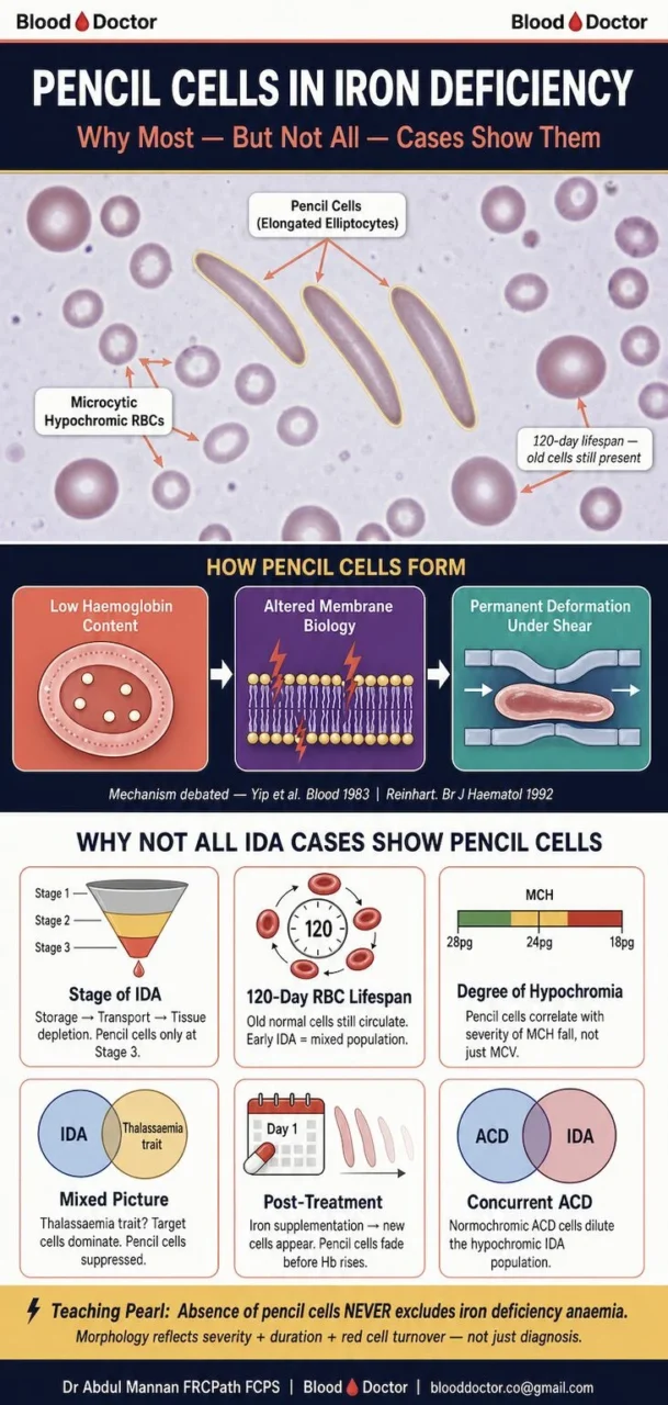
Pencil cells (elongated elliptocytes) appear in iron deficiency because low haemoglobin content changes how the red cell behaves under shear stress in the microcirculation. But the mechanism is genuinely debated.
Yip and Mohandas (Blood, 1983) found increased membrane stiffness from oxidative damage.
Reinhart (BJH, 1992) found the opposite — membrane elasticity is preserved and the cells actually pass more easily through narrow pores because of microcytosis.
Red cells live 120 days. Early iron deficiency means old, normally-shaped cells are still circulating. The film looks unremarkable. Biochemistry is already abnormal.
Severe hypochromia matters more than microcytosis alone. A patient with MCH of 18 pg shows pencil cells. One with MCH of 24 pg often does not.
Thalassaemia trait, anaemia of chronic disease, or partial treatment all suppress pencil cell formation even when IDA is present
Ford (Int J Lab Haematol, 2013) put it directly: the classic blood film appearances in iron deficiency are unreliable in practice.
So the teaching point is this.
Absence of pencil cells never excludes IDA. Their presence tells you something has been going on long enough, severely enough, for the circulating pool to turn over.
Stage. Severity. Duration. Concurrent conditions. That is why not all IDA films look the same.
What morphological finding has surprised you most in a confirmed IDA case?”
Other posts featuring Abdul Mannan on Hemostasis Today.
-
Sep 17, 2026, 20:38Konstantinos Bistas: Reduced Circulating Tregs in Pediatric Chronic Neutropenia
-
Sep 17, 2026, 15:33Gopal Patidar: Honoured to Contribute to the ASFA 2026 Guidelines for Therapeutic Apheresis
-
Sep 17, 2026, 20:36Cosima C. Hoch: Developing a 3D Organotypic Co-Culture Model for Pleomorphic Adenoma
-
Sep 17, 2026, 20:19Riten Kumar: Boston Children’s Hospital Ranked No. 1 in Seven Pediatric Specialties
-
Sep 17, 2026, 20:17Katrina Snow: Whole Blood or Component Therapy – Which Would You Choose?
-
Sep 17, 2026, 16:30Daria Camilli: Empowering Bleeding Disorder Advocacy Through Health Economics
-
Sep 17, 2026, 16:17Mehran Ghasemzadeh: Platelet NK Cell Crosstalk in Advanced Coronary Artery Disease
-
Sep 17, 2026, 16:16Barbara Adams Krolak: Do GLP-1s Affect Blood Clot Risk?
-
Sep 17, 2026, 15:46Heghine Khachatryan: Clinical Context at the Core of Thrombosis and Thrombophilia Management