


Aryabhatta Sadhu: Sickle Cell Crisis – When Diagnostics Should Change Therapy
Aryabhatta Sadhu, Attending Consultant and Head of Transfusion Medicine at Fortis Hospital Shalimar Bagh, New Delhi, shared a post on LinkedIn:
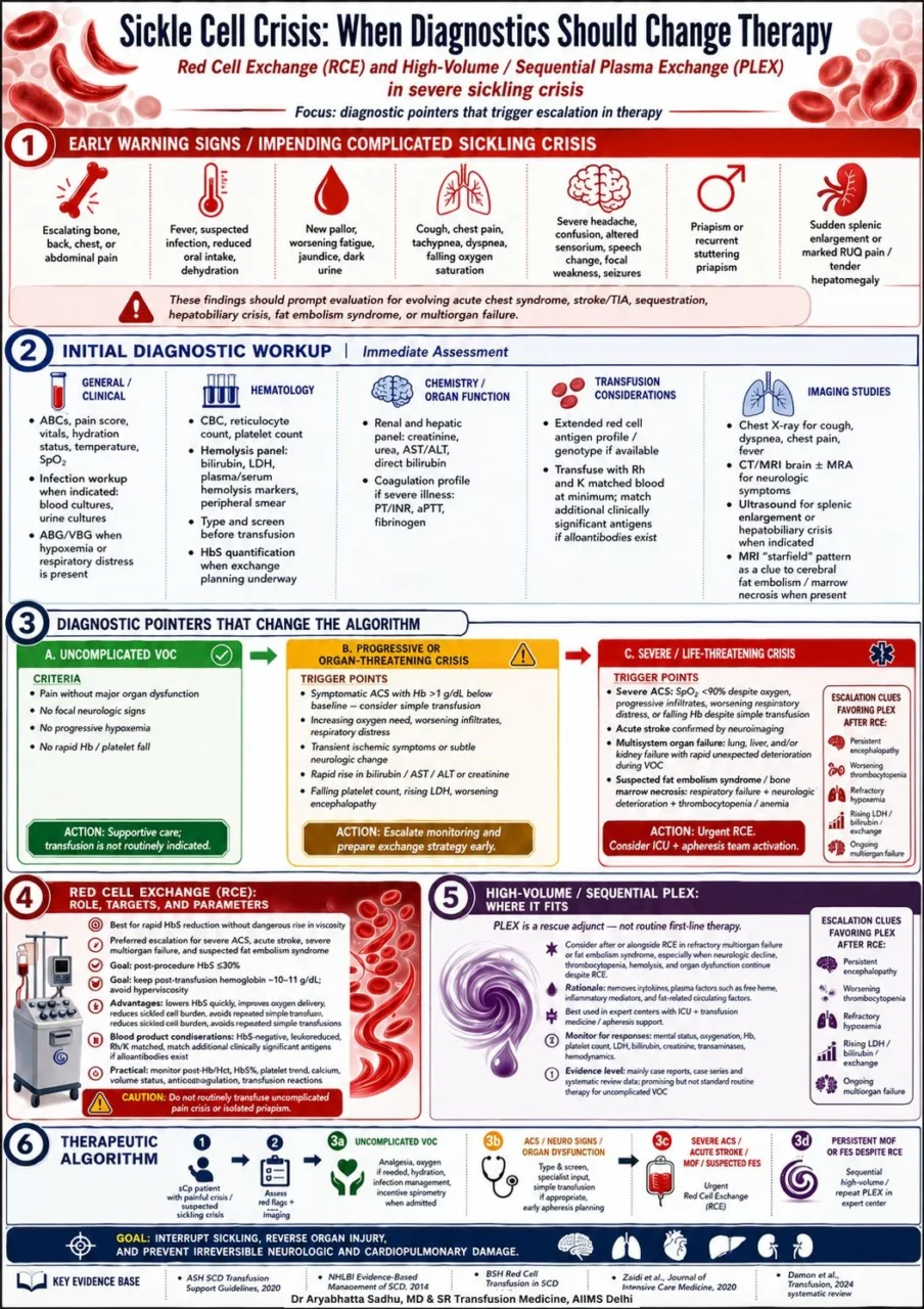
“The Dangerous Turn in Sickle Cell Crisis: Detect Early, Exchange Fast
Sickle cell crisis is not one clinical event.
It is a spectrum – from uncomplicated vaso-occlusive pain to acute chest syndrome, stroke, fat embolism syndrome, and multiorgan failure.
The real decision point is identifying when diagnostics should change therapy.
This infographic maps a practical escalation pathway for severe sickling crisis, focusing on:
- Early warning signs of impending complicated crisis
- Diagnostic triggers separating uncomplicated VOC from organ-threatening disease
- When simple transfusion may be sufficient
- When urgent red cell exchange is required
- Where high-volume / sequential plasma exchange may fit as rescue adjunct therapy in refractory multiorgan failure or fat embolism syndrome
Hard decision points for HbS% tracking:
- In uncomplicated VOC, HbS% alone should not trigger transfusion. Clinical deterioration matters more.
- In ACS, neurologic symptoms, falling SpO₂, progressive infiltrates, organ dysfunction, or suspected fat embolism syndrome – send HbS% early and activate transfusion/apheresis planning.
- In severe ACS, acute stroke, multiorgan failure, or suspected FES – do not wait for HbS% to return if the patient is clinically deteriorating. Start urgent red cell exchange planning.
- Post-RCE target: HbS less than or equal to 30%, with hemoglobin generally kept around 10–11 g/dL to avoid hyperviscosity.
- Persistent HbS more than 30% after RCE, or rebound HbS with worsening hypoxemia, encephalopathy, thrombocytopenia, rising LDH/bilirubin/creatinine, should trigger repeat clinical review, repeat HbS quantification, and consideration of further exchange or adjunct PLEX in expert settings.
The key point is simple:
Do not transfuse reflexively for uncomplicated pain crisis.
Do not delay exchange when hypoxemia, neurologic decline, organ failure, or fat embolism physiology appears.”
Other posts featuring Aryabhatta Sadhu with Hemsotasis Today.
-
Oct 12, 2026, 21:51Sam Botchey: Orthobiologics Are Advancing but the Evidence Must Keep Pace
-
Oct 12, 2026, 21:50Serum Vitamin D, Platelet Function, and Bleeding in Primary ITP – Platelets Journal
-
Oct 12, 2026, 21:30Nika Samadzadeh Tabrizi: Looking Forward to the PACeS Trial on Anticoagulation for POAF
-
Oct 12, 2026, 21:28Michael Megaly: Does Coronary Ectasia Affect Outcomes in Acute Coronary Syndromes?
-
Oct 12, 2026, 21:25Vitor Borin P. de Souza: Think Twice Before Anticoagulating an Isolated Subsegmental PE
-
Oct 12, 2026, 20:43Jazlyn A Selvasingh: Excited to Share Our Research at AAO-HNSF 2026 in Los Angeles
-
Oct 12, 2026, 20:31Natalia Kovtun: Higher Ferritin Cutoffs Now Define Iron Deficiency
-
Oct 12, 2026, 19:39Edward Loftus: Using Low-Dose Aspirin in Pregnant Women With IBD
-
Oct 12, 2026, 19:32Shruti Sachdeva: Strengthening the Fight against Anemia