

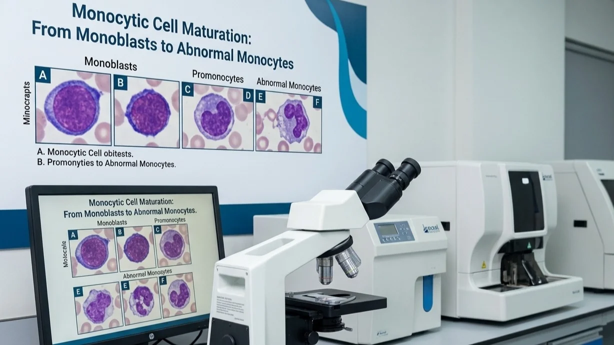
Saoud Hassan: Identifying Monocytic Precursors Under the Microscope
Saoud Hassan, Medical Laboratory Technologist at Excel Labs (Pvt) Ltd, shared a post on LinkedIn:
“Understanding Monocytic Cell Maturation: From Monoblasts to Abnormal Monocytes
Accurate identification of monocytic lineage cells is essential in hematology, especially when evaluating acute leukemias and myelodysplastic disorders.
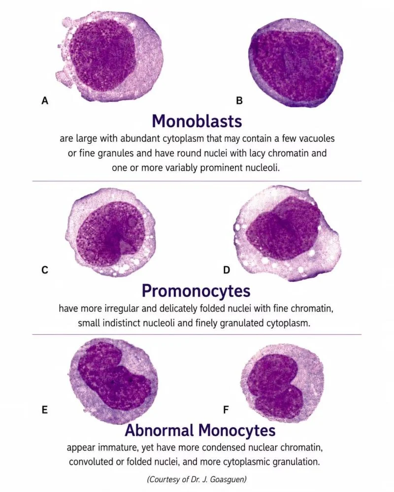
Monoblasts (A and B)
- Large immature cells with abundant basophilic cytoplasm.
- May contain fine granules or a few vacuoles.
- Round to oval nucleus with delicate (lacy) chromatin.
- One or more prominent nucleoli are usually visible.
Promonocytes (C and D)
- Intermediate stage between monoblasts and mature monocytes.
- Nucleus becomes more irregular, folded, or indented.
- Chromatin remains relatively fine.
- Nucleoli become less prominent.
- Cytoplasm shows fine granulation.
Abnormal Monocytes (E and F)
- May appear immature but demonstrate more condensed chromatin.
- Characterized by convoluted or folded nuclei.
- Increased cytoplasmic granulation compared to earlier precursors.
- Frequently observed in monocytic leukemias and other hematologic abnormalities.
Key Learning Point:
As monocytic cells mature, the nucleus changes from a round shape with delicate chromatin to a more folded and convoluted appearance, while chromatin condensation and cytoplasmic granulation progressively increase.
Understanding these subtle morphological differences is crucial for laboratory professionals, hematologists, and pathology trainees in making accurate diagnoses.”
Stay updated with Hemostasis Today.
-
Oct 19, 2026, 03:54Jennifer Trowbridge: How Mutant Blood Stem Cells Use Inflammation to Drive Their Own Expansion
-
Oct 19, 2026, 03:39Wyenona Hicks: Catching Up With Old Friends and New Colleagues at AABB 2026
-
Oct 19, 2026, 03:33Developing a Non-Invasive Treatment for Life-Threatening PE – University of Michigan College of Engineering
-
Oct 19, 2026, 03:28Jim Hoffman: How NETs Contribute to Gallstone Formation and Growth
-
Oct 19, 2026, 03:28Nicolas Hubacz: The Organized Chaos of Mitochondria
-
Oct 19, 2026, 03:02Neda Horvacki: Using ROTEM to Refine Transfusion Decisions in Acute Trauma
-
Oct 19, 2026, 02:45Diego Escarraman Martinez: MATTERs Study – When a Cohort Gave Us Major Clues about TXA in Trauma
-
Oct 19, 2026, 02:35Brian O Mahony: Promoting Physical Activity and Sport in Haemophilia Care
-
Oct 19, 2026, 02:35Malusi Mtetwa: Tranexamic Acid – What Does the Evidence Tell Us?