


Priyadarshini Ramakrishnan: Schistocytes – The Most “Misleading Heroes” in a Peripheral Smear
Priyadarshini Ramakrishnan, DHA-licensed Clinical Pathologist, shared a post on LinkedIn:
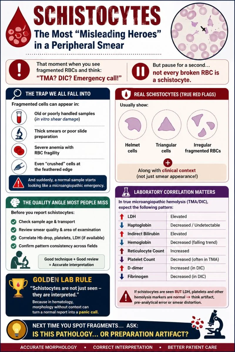
“Schistocytes: The most ‘misleading heroes’ in a peripheral smear
One glance at fragmented RBCs, and the immediate thought is often:
‘TMA? DIC? Is this an emergency?’
But here’s one of the biggest interpretation traps in hematology:
Not every fragmented red cell is a true schistocyte
Fragmented cells may be seen due to:
- Poor smear preparation
- Old or improperly handled samples (in vitro damage)
- Thick smears or feathered-edge artifacts
- Increased RBC fragility in severe anemia
Without careful interpretation, a perfectly routine sample can be mistaken for a microangiopathic emergency.
So, when should schistocytes raise concern?
True schistocytes should be accompanied by evidence of hemolysis, not just morphology.
Expected laboratory correlation:
- High LDH
- Low Haptoglobin
- High Indirect bilirubin
- Low Hemoglobin (falling trend)
- High Reticulocyte count
- Plus or minus low Platelet count (especially in thrombotic microangiopathies)
If the smear reports schistocytes but LDH and platelets are normal, with no biochemical evidence of hemolysis, pause before labeling it as microangiopathic hemolysis. Review the smear, assess pre-analytical factors, and correlate clinically.
A good laboratory professional doesn’t just identify schistocytes—they interpret them in context
Because morphology raises suspicion, but clinical and laboratory correlation confirms the diagnosis.
Have you ever encountered a case where an artifact nearly led to an overcall”
Stay updated with Hemostasis Today.
-
Oct 7, 2026, 16:44Amor Salah: 2026 NCS/SCCM Guidelines for Managing Antithrombotic-Associated ICH
-
Oct 7, 2026, 16:04NETs in Thrombosis: From Mechanism to Therapeutic Target
-
Oct 7, 2026, 15:28Amir Samimi: Prognostic Value of Platelet Count in Resected HNSCC
-
Oct 7, 2026, 15:21Eloïse Laouenan: Bridging Immunity and Thrombosis in MVTE
-
Oct 7, 2026, 15:10Uthra Sundaram: Honouring Donors During International Plasma Awareness Week
-
Oct 7, 2026, 15:04Daniel Victor Ortigoza: Should Frail Older Patients With AF Switch From VKA to DOACs?
-
Oct 7, 2026, 14:53Susan Noone: Donor Turned Recipient Shares Lifesaving Transfusion Story
-
Oct 7, 2026, 14:49Aimee Burtenshaw: The Impact of the Infected Blood Inquiry on Hospital Transfusion Laboratories
-
Oct 7, 2026, 14:41Vikash Jaiswal: Does Timing Matter for Calcium in Pre-Eclampsia Prevention?